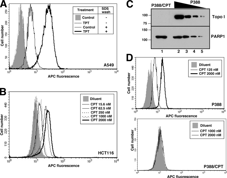
Figure 3.
Detection of topo I-DNA covalent complexes by flow cytometry. (A and B) A549 cells (A) or HCT116 cells (B) were incubated with 5 μM TPT (A) or the indicated TPT concentration (B) for 1 h, sedimented, lysed, fixed, stained with α-TopoIcc antibody followed by Alexa Fluor 647-conjugated secondary antibody and subjected to flow microfluorimetry. (C) Aliquots containing 50 µg of whole cell lysate from the P388/CPT and parental P388 mouse lymphoma lines (lanes 1 and 2) or serial 2-fold dilutions (lanes 3-5) were subjected to SDS-PAGE, probed for topo I and, as a loading control, poly(ADP-ribose) polymerase 1 (PARP1). (D) Detection of topo I-DNA covalent complexes in P388 (top) and P388/CPT cells (bottom) by flow microfluorimetry.

