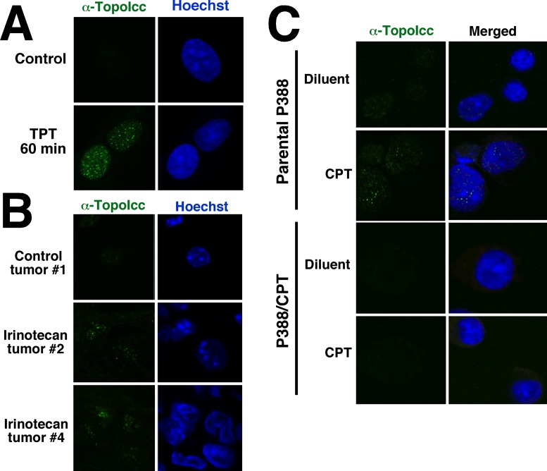
Figure 4.
Detection of topo I-DNA covalent complexes by fluorescence microscopy. (A) After treatment for 1 h with 1 μM TPT, A549 cells were fixed, permeabilized, incubated with SDS and stained with α-TopoIcc antibody (green) and Hoechst 33258 (blue). (B) Cryostat sections of A549 xenografts harvested 0 (top), 2 (middle) and 4 h (bottom) after irinotecan administration were stained with α-TopoIcc and Hoechst 33258. (C) After treatment of P388 or P388/CPT cells with 1 μM camptothecin for 30 min, cells were prepared and stained with α-TopoIcc.

