Figure 4.
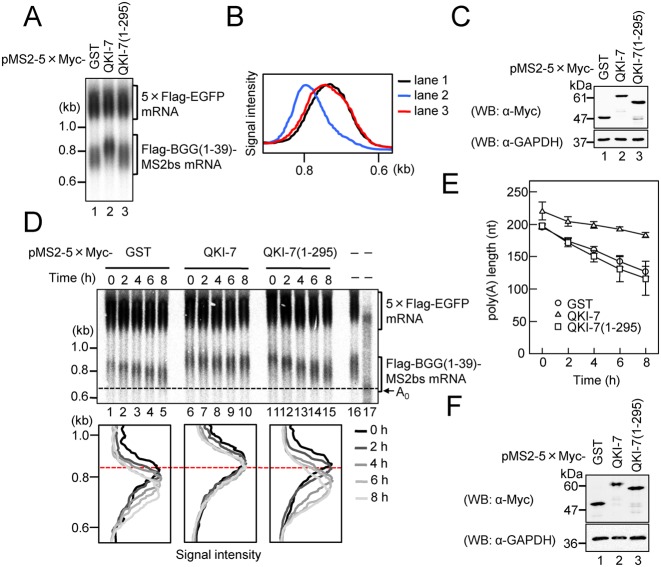
The C-terminal region of QKI-7 is necessary for induction of polyadenylation. (A) HEK293T cells were co-transfected with the pFlag-CMV5/TO-BGG(1–39)-MS2bs reporter plasmid, the pCMV-5×Flag-EGFP reference plasmid and either pMS2–5×Myc-GST, pMS2–5×Myc-QKI-7, pMS2–5×Myc-QKI7(1–295). BGG(1–39)-MS2bs mRNA was detected by Northern blot analysis. (B) BGG(1–39)-MS2bs mRNA length distribution was visualized as in Figure 1D. (C) Protein expression was analyzed by Western blotting with the indicated antibodies. (D) HEK293T cells were co-transfected with the pFlag-CMV5/TO-BGG (1–39)-MS2bs reporter plasmid, the pCMV-5×Flag-EGFP reference plasmid, the pT7-TR plasmid and either pMS2–5×Myc-GST (lanes 1–5), pMS2–5×Myc-QKI7 (lanes 6–10) or pMS2–5×Myc-QKI7 (1–295) (lanes 11–15). One day later, synthesis of the BGG(1–39)-MS2bs mRNA was induced by treatment with tetracycline for 2 h. Cells were harvested at the specified times after BGG(1–39)-MS2bs mRNA production was shut off. (Upper panel) BGG(1–39)-MS2bs mRNA was detected by Northern blot analysis. To mark deadenylated (A0) BGG(1–39)-MS2bs mRNA, steady-state BGG(1–39)-MS2bs mRNA (lane 16) was digested with RNase H in the presence of oligo(dT) (lane 17). (Lower panel) BGG(1–39)-MS2bs mRNA length-distribution was visualized as in Figure 1D. (E) The average length of poly(A) tails at each time point was measured as in Figure 2B. Error bars represent the standard deviation of three independent experiments. (F) Protein expression was analyzed by Western blotting with the indicated antibodies.

