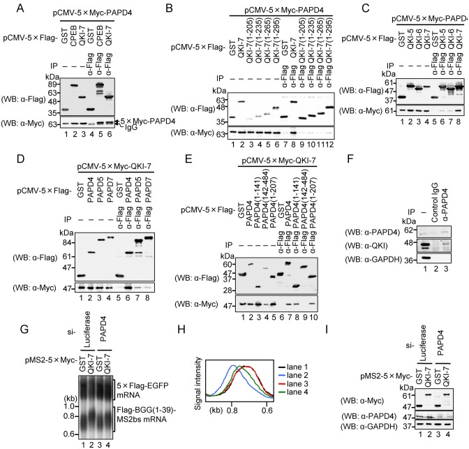
Figure 5.
QKI-7 specifically interacts with PAPD4 through its unique C-terminal region to induce mRNA polyadenylation. (A) HEK293T cells were co-transfected with pCMV-5×Myc-PAPD4 and either pCMV-5×Flag-GST (lanes 1 and 4), pCMV-5×Flag-CPEB (lanes 2 and 5) or pCMV-5×Flag-QKI-7 (lanes 3 and 6). Cell extracts were subjected to immunoprecipitation (IP) using the anti-Flag antibody. The immunoprecipitates (lanes 4–6) and inputs (lanes 1–3) were analyzed by Western blotting with the indicated antibodies. (B) HEK293T cells were co-transfected with pCMV-5×Myc-PAPD4 and either pCMV-5×Flag-GST (lanes 1 and 7), pCMV-5×Flag-QKI-7 (lanes 2 and 8), pCMV-5×Flag-QKI-7(1–205) (lanes 3 and 9), pCMV-5×Flag-QKI-7(1–235) (lanes 4 and 10), pCMV-5×Flag-QKI-7(1–265) (lanes 5 and 11) or pCMV-5×Flag-QKI-7(1–295) (lanes 6 and 12). Cell extracts were subjected to IP using the anti-Flag antibody. The immunoprecipitates (lanes 7–12) and inputs (lanes 1–6) were analyzed by Western blotting with the indicated antibodies. (C) HEK293T cells were co-transfected with pCMV-5×Myc-PAPD4 and either pCMV-5×Flag-GST (lanes 1 and 5), pCMV-5×Flag-QKI-5 (lanes 2 and 6), pCMV-5×Flag-QKI-6 (lanes 3 and 7) or pCMV-5×Flag-QKI-7 (lanes 4 and 8). Cell extracts were subjected to IP using the anti-Flag antibody. Immunoprecipitates (lanes 5–8) and inputs (lanes 1–4) were analyzed by Western blotting with the indicated antibodies. (D) HEK293T cells were co-transfected with pCMV-5×Myc-QKI-7 and either pCMV-5×Flag-GST (lanes 1 and 5), pCMV-5×Flag-PAPD4 (lanes 2 and 6), pCMV-5×Flag-PAPD5 (lanes 3 and 7) or pCMV-5×Flag-PAPD7 (lanes 4 and 8). Cell extracts were subjected to IP using anti-Flag antibody. Immunoprecipitates (lanes 5–8) and inputs (lanes 1–4) were analyzed by Western blotting with the indicated antibodies. (E) HEK293T cells were co-transfected with pCMV-5×Myc-QKI-7 and either pCMV-5×Flag-GST (lanes 1 and 6), pCMV-5×Flag-PAPD4 (lanes 2 and 7), pCMV-5×Flag-PAPD4(1–141) (lanes 3 and 8), pCMV-5×Flag-PAPD4(142–484) (lanes 4 and 9) or pCMV-5×Flag-PAPD4(1–207) (lanes 5 and 10). Cell extracts were subjected to IP using anti-Flag antibody. Immunoprecipitates (lanes 6–10) and inputs (lanes 1–5) were analyzed by western blotting with the indicated antibodies. (F) HEK293T cell extracts were subjected to IP using anti-PAPD4 or normal goat IgG as a non-specific control. The immunoprecipitaes (lanes 2 and 3) and input (lane 1) were analyzed by Western blotting with the indicated antibodies. (G) HEK293T cells were co-transfected with the pFlag-CMV5/TO-BGG (1–39)-MS2bs reporter plasmid, the pCMV-5×Flag-EGFP reference plasmid, either pMS2–5×Myc-GST (lanes 1 and 3) or pMS2–5×Myc-QKI7 (lanes 2 and 4), and either Luciferase siRNA (lanes 1 and 2) or PAPD4 siRNA (lane 3 and 4). BGG(1–39)-MS2bs mRNA was detected by Northern blot analysis. (H) BGG(1–39)-MS2bs mRNA length-distribution was visualized as in Figure 1D. (I) Protein expression was analyzed by Western blotting with the indicated antibodies.

