Figure 6.
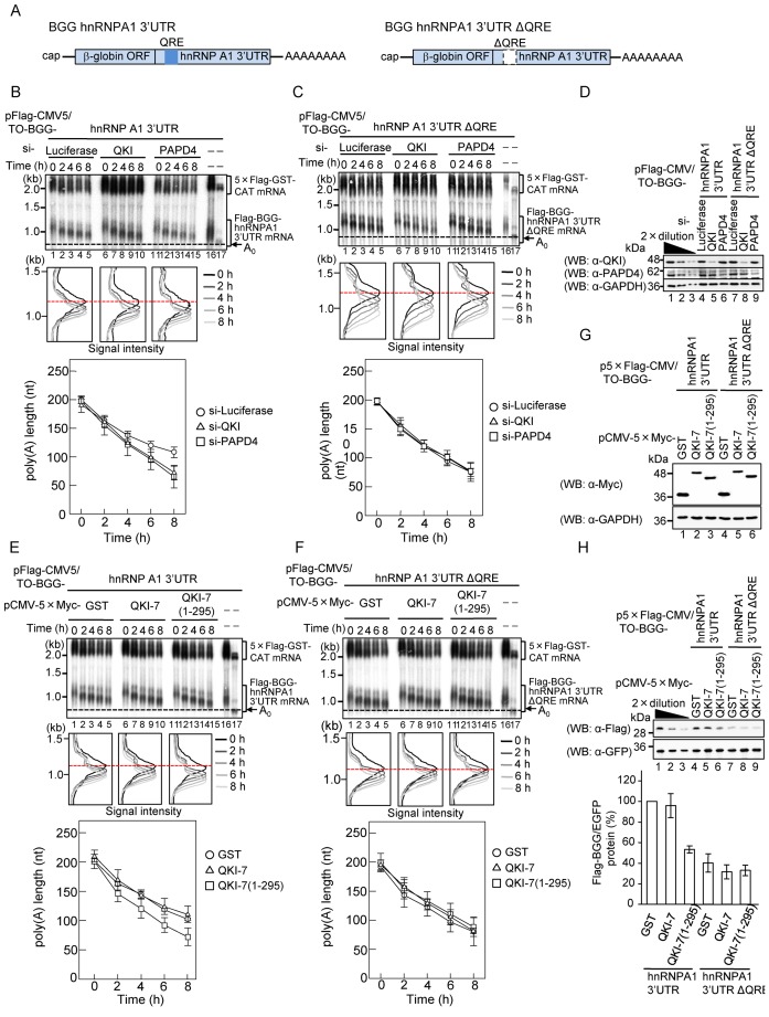
QKI and PAPD4 regulate the poly (A) tail length of hnRNPA1 mRNA in a QRE-dependent manner. (A) Schematic representation of BGG hnRNPA1 reporter constructs. The β-globin ORF (BGG) was appended with either the hnRNPA1 3′ UTR, which contained the QRE, or the hnRNPA1 3′ UTR in which the QRE had been deleted. (B and C) HEK293T cells were transfected with siRNA against luciferase (lanes 1–5), QKI (lanes 6–10) or PAPD4 (lanes 11–15). Cells were co-transfected with either pFlag-CMV5/TO-BGG-hnRNPA1 3′ UTR (Figure 6B), or pFlag-CMV5/TO-BGG-hnRNPA1 3′ UTR ΔQRE (Figure 6C) pCMV-5×Flag-GST-CAT reference plasmid and pT7-TR, 24 h after siRNA transfection. Pulse-chase analysis was performed as in Figure 4D. (Upper panel) BGG-hnRNPA1 3′ UTR mRNA was detected by Northern blot analysis. (Middle panel) BGG-hnRNPA1 3′ UTR mRNA length-distribution was visualized as in Figure 1D. (Lower panel) The average length of poly(A) tails at each time point was measured from the middle panel as in Figure 2B. Error bars represent the standard deviation of three independent experiments. (D) Protein expression was analyzed by Western blotting with the indicated antibodies. (E and F) HEK29T cells were co-transfected with either the pFlag-CMV5/TO-BGG-hnRNPA1 3′ UTR (Figure 6E) or the pFlag-CMV5/TO-BGG-hnRNPA1 3′ UTR ΔQRE (Figure 6F), the pCMV-5×Flag-GST-CAT reference plasmid, pT7-TR and either pCMV-5×Myc-GST (lanes 1–5), pCMV-5×Myc-QKI-7 (lanes 6–10) or pCMV-5×Myc-QKI-7(1–295) (lanes 11–15). Pulse-chase analysis was performed as in Figure 4D. (Upper panel) BGG-hnRNPA1 3′ UTR mRNA was detected by Northern blot analysis. (Middle panel) BGG-hnRNPA1 3′ UTR mRNA length-distribution was visualized as in Figure 1D. (Lower Panel) The average length of poly(A) tails at each time point was measured from the middle panel as in Figure 2B. Error bars represent the standard deviation of three independent experiments. (G) Protein expression was analyzed by Western blotting with the indicated antibodies. (H) HEK293T cells were co-transfected with either the p5×Flag-CMV5/TO-BGG-hnRNPA1 3′ UTR or the p5×Flag- CMV5/TO-BGG-hnRNPA1 3′ UTR ΔQRE, the pEGFP-C1 reference plasmid and either pCMV-5×Myc-GST, pCMV-5×Myc-QKI-7 or pCMV-5×Myc-QKI-7(1–295). Protein expression was analyzed by Western blotting with the indicated antibodies (upper panel). The amount of Flag-BGG protein was measured and normalized to the EGFP protein. The protein level in pCMV-5×Myc-GST transfected cells was defined as 100% (lower panel). The results were derived from three independent experiments and shown as the means ± SD.

