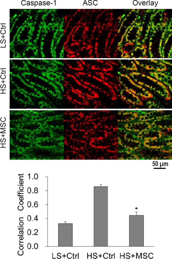
Fig. 4.
Confocal imaging for the colocalization of ASC and caspase-1 immunostainings in the renal medulla of Dahl S rats. Top: representative photomicrographs showing the immunostaining of caspase-1 (green) and ASC (red) as well as the overlaid images. Yellow color in the overlaid images represents the colocalization of caspase-1 and ASC. Bottom: summarized data showing the colocalization coefficient of caspase-1 and ASC immunostainings analyzed using a computer software Image-Pro Plus. *P < 0.05 vs. other groups, n = 4.

