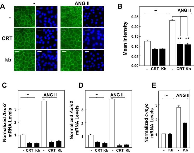
Fig. 2.
Protein kinase D (PKD) family inhibitors prevent β-Cat nuclear translocation and expression of Axin2 and c-myc in response to ANG II in intestinal epithelial cells. A: confluent cultures of IEC-18 cells were incubated in the absence (−) or presence of 2.5 μM CRT006610 (CRT) or 3.5 μM kb NB 142-70 (kb) for 1 h before stimulation of the cells without (−) or with 50 nM ANG II for 4 h. Then the cultures were fixed with 4% paraformaldehyde and stained with an antibody that detects β-Cat conjugated to Alexa Fluor 488 and with Hoechst 33342 to visualize the cell nuclei. B: quantification of β-Cat nuclear localization was determined with the CellProfiler software, as described in materials and methods and Fig. 1. Results shown here are the mean nuclear intensities ± SE (n = 1,500 cells) from 1 experiment, **P < 0.001. Similar results were obtained in 3 separate biological replicates. C and D: confluent cultures of IEC-18 cells were incubated in the absence or presence of CRT or kb for 1 h before stimulation of the cells without (−) or with 50 nM ANG II for either 1 h (C) or 2 h (D), as indicated. RNA was isolated, and the relative levels (n = 3) of Axin2 mRNA compared with GAPDH mRNA were measured by quantitative RT-PCR. E: confluent IEC-18 cells were stimulated with ANG II (50 nM) for 2 h. RNA was isolated, and the relative levels (n = 3) of c-myc mRNA compared with GAPDH mRNA were measured by quantitative RT-PCR. Similar results were obtained in separate experiment. Scale bars = 30 μm.

