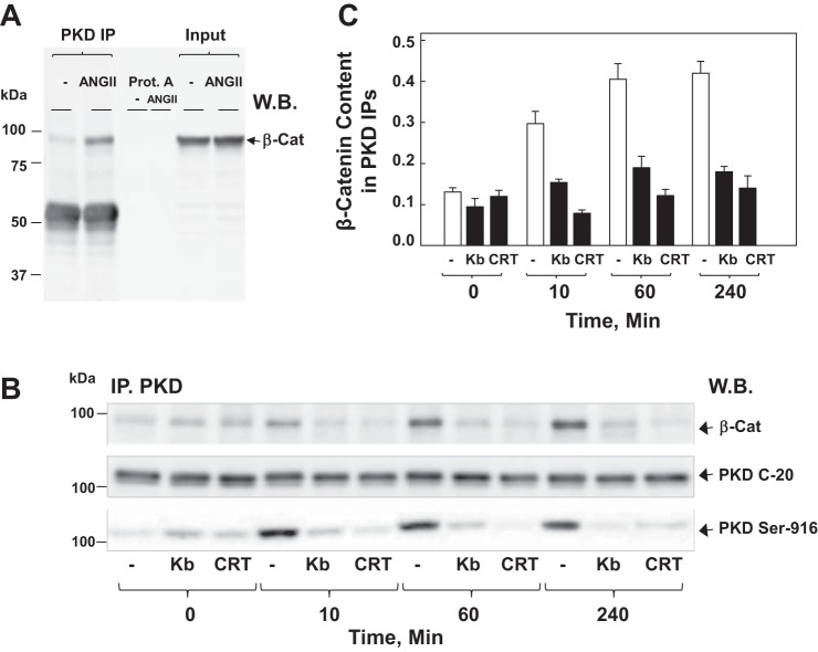
Fig. 7.
ANG II induces complex formation between β-Cat and PKD1 in IEC-18 cells. A: confluent cultures (100 mm dish/treatment, 6 × 10⁶ cells) of IEC-18 cells were stimulated with 50 nM ANG II for 60 min. Then the cells were lysed, and PKD C-20 immunoprecipitates (PKD IP) and protein A agarose negative controls (omitting the PKD C-20 antibody) were analyzed by Western blotting (W.B.) with antibodies that detect β-Cat. The input represents 1/30 of the total lysate used for IP. B: confluent cultures of IEC-18 cells were incubated in the absence (−) or presence (kb) of 3.5 μM kb or 2.5 μM CRT for 1 h before stimulation of the cells with 50 nM ANG II for either 10, 60, or 240 min, as indicated. Cells were lysed, and PKD IP were analyzed by W.B. with antibodies that detect β-Cat, PKD1 (PKD C-20), or PKD1 pSer916 (PKD-Ser916). C: bars represent the means ± SE obtained in 3 independent experiments. Individual values are the level of β-Cat band intensity normalized to the corresponding PKD band intensity in each experiment.

