Fig. 8.
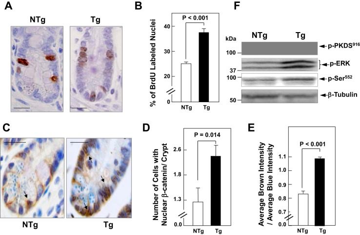
PKD1 signaling induces β-Cat nuclear translocation in intestinal crypt epithelial cells. A: the proportion of crypt cell DNA synthesis was determined by immunohistochemical detection of 5-bromo-2′-deoxyuridine (BrdU) incorporated into the nuclei of crypt cells of the ileum. Transgenic (Tg) mice and nontransgenic (NTg) littermates were injected intraperitoneally with BrdU (100 μg/g body wt) and killed 3 h after the injection. Tissue fixation, histological sections, and routine staining were performed as described in materials and methods. Scale bars = 20 μm. B: the bars represent the percentage of BrdU-labeled crypt cells (means ± SE) in the PKD1 Tg mice (solid bar) and in the NTg littermates in Tg mice (open bar). C: immunohistochemistry of histological sections from PKD1 Tg mice and NTg littermates with β-Cat (6B3) rabbit monoclonal antibody. Scale bars = 20 μm. D: the bars represent the number of cells with nuclear β-Cat in well-oriented crypts of PKD1 Tg mice and NTg littermates. Values are means ± SE (n = 8). E: the bars represent the ratio of brown intensity to blue intensity (β-Cat/nuclear) staining in crypt cells of PKD1 Tg compared with NTg. Values are means ± SE (n = 600 cells in Tg mice and n = 536 cells in NTg mice). F: epithelial cells from the ileum of Tg mice and NTg littermates mice were isolated sequentially by timed incubations in a EDTA-PBS solution. W.B. was used to analyze lysates of these cells for PKD1 autophosphorylated at Ser916 (pPKDS916), ERK at Thr202 and Tyr204 (pERK), and β-Cat at Ser552 (pSer552). Equivalent loading was verified by immunoblotting for tubulin.

