Fig. 2.
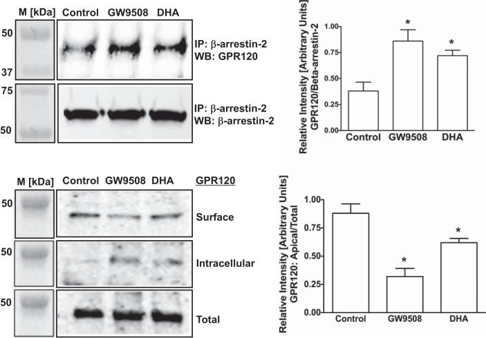
Agonist stimulation internalizes GPR120 via interaction with β-arrestin-2 in Caco-2 cells. Top left: lysates of control or agonist [50 μM GW9508 or 50 μM docosahexaenoic acid (DHA)]-treated (30 min) Caco-2 cells, containing equal amounts of proteins, were used to immunoprecipitate (IP) GPR120 with anti-β-arrestin-2 antibody. IPs were subjected to SDS-PAGE and probed with anti-GPR120 antibody in immunoblotting. After being stripped with 0.2 N NaOH, blots were reprobed with anti-β-arrestin-2 antibody. Representative blot of 3 independent experiments is shown. Top right: densitometric analysis of band intensities of GPR120/β-arrestin-2 in different groups is shown. Values are means ± SE; n = 3. *Different from control, P ≤ 0.05. Bottom left: monolayers of Caco-2. Ten-day postplating were treated with 50 μM GW9508 or 50 μM DHA for 30 min, and cell surface levels of GPR120 were measured by cell-surface biotinylation. Representative blot of 3 independent experiments is shown. Bottom right: densitometric analysis of band intensities of apical GPR120/total GPR120 in different groups is shown. Values are means ± SE; n = 3. *Different from control, P ≤ 0.05. WB, Western blot.

