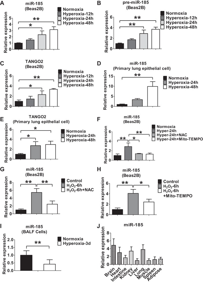
Fig. 1.
Oxidative stress or hyperoxia induces miR-185 expression in lung epithelial cells. A and B: time course of hyperoxia-induced expressions of miR-185 (A), miR-185 precursor (B), and TANGO2 (C) in Beas2B cells detected using real-time PCR. D and E: primary lung epithelial cells isolated from mice were exposed to hyperoxia for the indicated time points. The expression of miR-185 (D) and TANGO2 (E) in mouse primary lung epithelial cells was determined using real-time PCR. F: Beas2B cells were exposed to normoxia or hyperoxia for 24 h in the presence or absence of N-acetyl-l-cysteine (NAC) (5 mM) or Mito-TEMPO (100 μM). Real-time PCR was performed to determine the level of miR-185. G: Beas2B cells were treated with H2O2 (90 μM) for 6 h in the presence or absence of NAC (5 mM). The expression level of miR-185 was measured using real-time PCR. H: Beas2B cells were treated with H2O2 (90 μM) for 6 h in the presence or absence of Mito-TEMPO (100 μM). The expression level of miR-185 was measured using real-time PCR. I: real-time PCR analysis of miR-185 in mouse bronchoalveolar lavage fluid (BALF) exposed to normoxia (room air) or hyperoxia (3 days, n = 6 for each group). J: real-time PCR analysis of miR-185 in different mouse tissues (n = 3). All the data are representative of 3 independent experiments. *P < 0.05, **P < 0.01.

