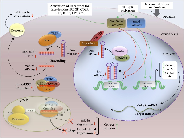
Fig. 1.
Biogenesis of miRNA 29a and its regulation of Col3A1 mRNA inside an activated fibroblast in scleroderma. Schematics illustrate the transcription of pri-miR-29a being regulated through TGF-β, PDGF, CTGF, ET-1, IL-4, and LPS signaling via SMAD and non-SMAD pathways, and by epigenetic silencing. Transcription of Col3A1 mRNA is regulated by the overactive SMAD and non-SMAD signaling pathways. Extracellular exchange of miR 29a via exosomes (small vesicles produced by budding of plasma membrane) is consequently reduced, resulting in low circulating miR 29a levels in the very early stage of scleroderma, which could potentially serve as a disease biomarker (60). In human beings, imperfect complementarity of miR-mRNA binding is more common than perfect complementarity; therefore, miR-induced translational repression (thick black line) is more common that target mRNA degradation (thin black line). *Mechanical stress also activates fibroblasts to synthesize excess collagen (34). Red lines and arrows indicate inhibition or decrease; green lines and arrows indicate stimulation or increase. AGO2, Argonaute2; CTGF, connective tissue growth factor; DGCR8, DiGeorge syndrome critical region gene 8; ET-1, endothelin-1; IGF-1, insulin like growth factor-1; IL-4, interleukin-4; LPS, lipopolysaccharide; ORF, open reading frame; P body, processing body; PDGF-β, platelet-derived growth factor-β; Pre-miR, precursor miR; Pri-miR, primary miR; RAN-GTP, RAS-related nuclear protein-guanosine triphosphate; RISC, RNA-induced silencing complex; TRBP, transactivating response RNA-binding protein; TGF-β, transforming growth factor-β; UTR, untranslated region.

