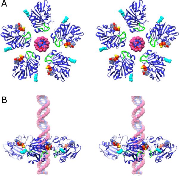
Figure 5.

Ring structure of the gp16 and DNA. A) Cross-eyed stereo diagram of the ring structure of gp16 based on fitting the X-ray structure of the monomer into its corresponding cryoEM density in a 5-fold averaged reconstruction of particles stalled during packaging via the addition of γ-S-ATP. The viewing direction is from outside the phage looking toward the interior. The putative DNA translocating loop is shown in green, the arginine finger R146 is shown as cyan van der Waal spheres, and the substrate analog AMP-PNP and DNA as van der Waal spheres colored by element. B) Stereo-diagram of a side-view of the gp16 ring, colored as in (A); the capsid would be above the ring, and the back-half of the ring has been left out to facilitate viewing.
