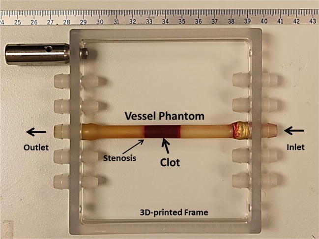
Figure 2.

The vessel phantom is held by a 3D-printed frame and can be connected in line with the flow model using tubing fittings. A 35% stenosis is located in the vessel phantom to fix the clot formed to one side so that it does not slip under pressure. The inner diameter is 4.2 mm on the left of the stenosis and 6.5 mm on the right. A clot is formed on the right side of the stenosis as shown in the figure.
