Figure 6.

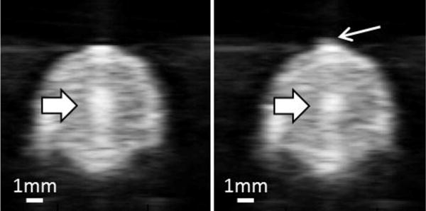
Ultrasound images of vessel lumen during treatment. Left: Focal cavitation at the center of the vessel lumen only (block arrow). Right: Focal cavitation (block arrow) with weak pre-focal cavitation (line arrow).

Ultrasound images of vessel lumen during treatment. Left: Focal cavitation at the center of the vessel lumen only (block arrow). Right: Focal cavitation (block arrow) with weak pre-focal cavitation (line arrow).