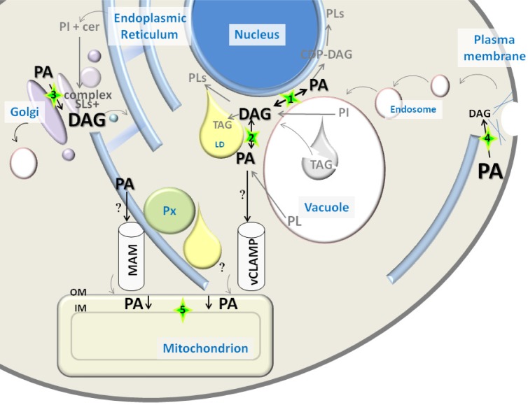
Figure 1.
PA and DAG distribution in yeast. PA and DAG font size reflects abundance of pools of these lipids in different cellular compartments, based on data obtained by combined subcellular fractionation and lipidomics approaches (Table 2). Relevant contact sites between organelles for PA/DAG conversion or transport are highlighted with a green star. Arrows involved in PA/DAG conversions reflect the presence of associated enzymatic activities in that compartment: (1) Nuclear vacuolar junction, presence of PA phosphatase Pah1 and endoplasmic reticulum (ER) localized Dgk1; (2) Vacuole, presence of PA phosphatase Dpp1 and phospholipases. It is not clear if Dgk1 localizes to the vacuole; (3) Golgi, presence of PA phosphatase Lpp1. Large pool of DAG is produced by complex sphingolipid biosynthesis; (4) Plasma membrane/actin patches (blue lines), presence of PA phosphatase App1; (5) PA transport into mitochondria is mediated by Ups1-Mdm35. Putative sources of mitochondrial PA are denoted by a question mark.
Abbreviations: CDP-DAG, CDP-diacylglycerol; Cer, ceramide; DAG, diacylglycerol; ER, endoplasmic reticulum; IM, inner membrane; LD, lipid droplet; MAM, mitochondria-associated membrane; OM, outer membrane; PA, phosphatidic acid; PI, phosphatidylinositol; PL, phospholipid; Px, peroxisome; SLs, sphingolipids; TAG, triacylglycerol; vCLAMP, vacuole and mitochondria patch.

