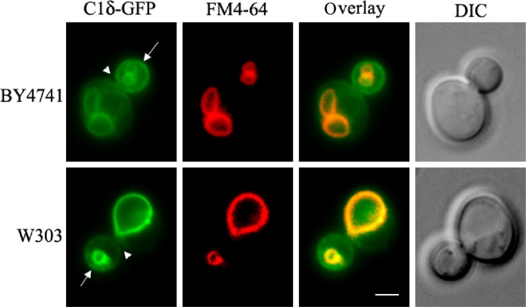
Figure 2.
C1δ–GFP localization in wild-type yeast. The tandem C1A–C1B domains of PKCδ (Table 2) fused to GFP (C1δ–GFP)102 were cloned into the p416-GPD (CEN, URA3) vector for constitutive expression in yeast. Wild-type cells (BY4741 and W303-1A) carrying this plasmid were grown to mid-log phase in synthetic defined selective medium at 30°C. For vacuolar membrane staining, cells were incubated with 16 μM FM4-64 (Molecular Probes®) for 15 minutes at 30°C and allowed to grow for an additional 30 minutes before the preparation of slides for imaging immediately after. Cells were imaged live using a Zeiss Imager Z.1 epifluorescence microscope. Arrows point to the nonvacuolar GFP signal detected in the plasma membrane of the bud, while arrowheads point to the bud neck. Scale bar = 2 μm.
Abbreviation: DIC, differential interference contrast.

