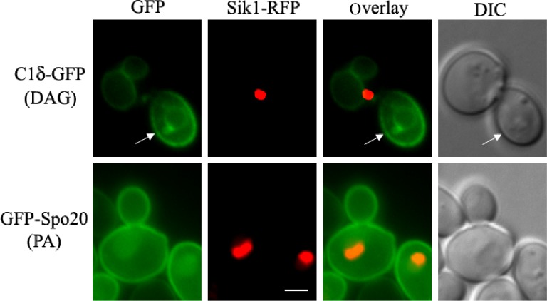
Figure 3.
Comparison of PA and DAG distribution in yeast. Wild-type yeast (BY4741) cells expressing the nucleolar protein Sik1 fused to RFP from the endogenous locus104 was transformed with plasmids carrying C1δ–GFP (CEN, URA3) or GFP–Spo20[51–91] (2 μ, URA3; kindly provided by Neiman et al)73 for the detection of DAG or PA, respectively. Transformants were grown to mid-log phase in synthetic defined selective medium at 30°C. Cells were imaged live using a Zeiss Imager Z.1 epifluorescence microscope. Arrows point to the nonvacuolar GFP signal detected in the plasma membrane of the bud. Scale bar = 2 μm.
Abbreviation: DIC, differential interference contrast.

