
Figure 2.
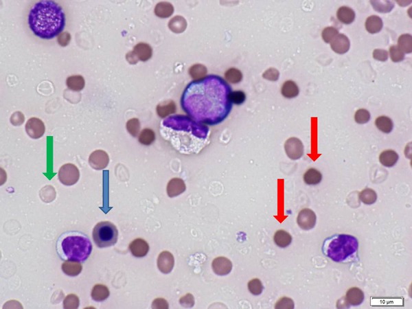
Peripheral blood smear using the sample collected in the ER of the local hospital. Note that there is marked anisocytosis (red cells of varying sizes), spherocytosis (red arrows), nucleated red blood cells (blue arrows), and dehemoglobinized ghost cells (green arrow).