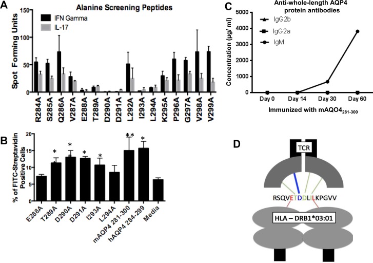
Fig 4. Identification of critical residues of human (h)AQP4281-300 for presentation in the context of HLA-DRB1*03:01 and recognition by the B.10 T cell receptor (TCR).
(A) First, the ability of hAQP4281-300-reactive lymph node cells to recognize the alanine screening peptides was determined by ELISpot. 5.0x10⁵ cells/well lymph node cells taken ten days post immunization of HLA-DRB1*03:01 transgenic mice with hAQP4281-300 were restimulated with hAQP4 alanine scanning peptides (2 5 μg/mL) for 48 hours in IFNγ and IL-17 ELISpot plates (* = P-value < 0.05 and ** = P-value < 0.01). (B) Alanine screening peptides that not result in an increased frequency of IFNγ and IL-17 secreting lymph node cells were identified as the key residue peptides, and were subsequently tested in a MHC binding assay. Splenocytes taken from HLA-DRB1*03:01 transgenic mice were incubated for 12 hours in the presence of biotinylated hAQP4 alanine scanning peptides. Post incubation, cells were stained utilizing FITC-Avidin, and antigen positive cells were quantified by flow cytometry (* = P-value < 0.05 and ** = P-value < 0.01). (C) There was no Ig isotype class switch in mice immunized with mAQP4284-299 with regard to antibody responses against whole-length AQP4 protein. (D) Critical HLA-DRB1*03:01 anchor residues, and B.10 TCR contact amino acids are specified. E288 and L294 are required as HLA-DRB1*03:01 anchor residues, while T289, D290, D291, and I293 are critical B.10 TCR interacting residues.

