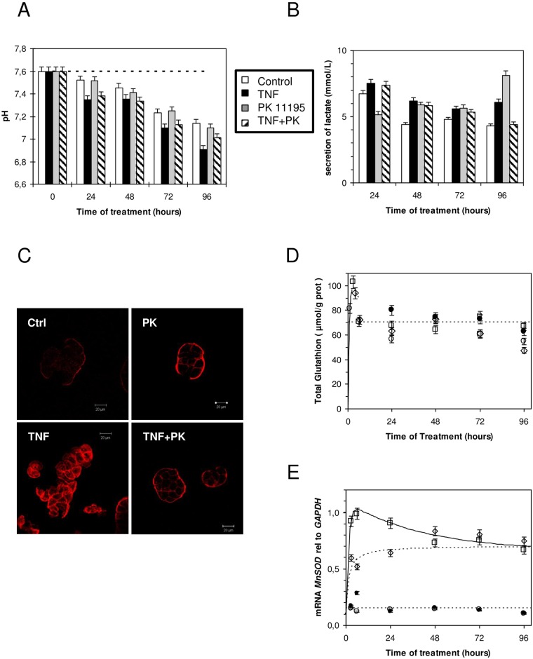
Fig 3. HT-29 cell metabolism upon TNF treatment.
(A) Time course of cell culture medium pH change as a function of treatment (open bars, control; black bars, 10 ng/mL of TNF renewed daily; grey bars, 1 μM of PK 11195 treatment; dashed bars, 10 ng/mL of TNF and 1 μM of PK 11195, renewed daily). (B) Time-dependent lactate accumulation in cell culture medium; treatment as in panel A. (C) Actin stress fiber formation after 4 hours upon similar treatment as in panel A (control, upper left panel; 1 μM of PK 11195-treated cells, upper right panel; 10 ng/mL of TNF-treated cells, lower left panel; 10 ng/mL of TNF and 1 μM of PK 11195-treated cells, lower right panel). (D) Time-dependent total glutathione measurement; treatment as in panel A (closed circles, control; open squares, in the presence of 10 ng/mL of TNF daily replaced; grey triangles, 1 μM of PK 11195 replaced daily; grey diamonds, in the presence of 10 ng/mL of TNF and 1 μM of PK 11195, replaced daily). (E) Time course of MnSOD mRNA expression relative to GAPDH; treatment as in panel A (open circles, control; closed circles, in the presence of 1 μM of PK 11195, replaced daily; Open squares, in the presence of 10 ng/mL of TNF, replaced daily; open diamonds, in the presence of 10 ng/mL of TNF and 1 μM of PK 11195, replaced daily). The results are expressed as the means ± standard error of the mean.

