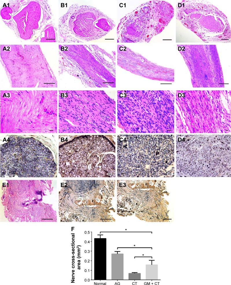
Figure 6.
Histology of regenerated nerves for HE staining and Loyez staining under light microscopy at 20 weeks postoperation.
Notes: (A) Normal group. (B) AG group. (C) CT group. (D) GM + CT group. (E) BLANK group. (A1–D1) HE staining of transverse sections of midsection of regeneration nerve. (A2–D2, A3–D3, and E1) HE staining of longitudinal histology of regenerated nerves. (A4–D4 and E2, E3) Loyez staining. (E3) Neuroma formed at the end of the proximal stump (bar in [A1–D1] and [E3] =200 μm; bar in [A2–D2] and [E1 and E2] =500 μm; bar in [A3–D3] =20 μm; bar in [A4–D4] =10 μm). (F) Statistical analysis of the nerve cross-sectional area *P<0.05.
Abbreviations: HE, hematoxylin–eosin; AG, autograft; CT, collagen tube; GM + CT, collagen tube filled with GDNF-loaded microspheres; GDNF, glial cell-line derived neurotrophic factor.

