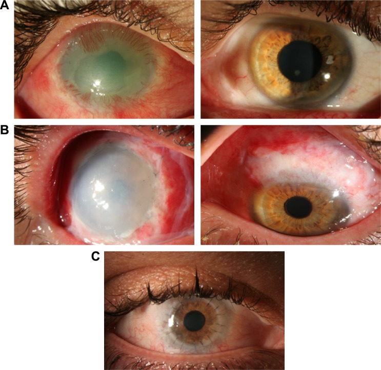
Figure 3.
Pre- and postautologous stem cell transplantation photographs.
Notes: (A) Patient with limbal stem cell deficiency after chemical burn injury, with neovascularization and scarring (left) and donor eye with healthy stem cell niche (right). (B) Slit-lamp photograph after autologous stem cell transplant in affected (left) and donor eye (right) with evident wide excision. (C) Slit-lamp photograph after penetrating keratoplasty one year after initial stem cell transplant.

