Figure 2.
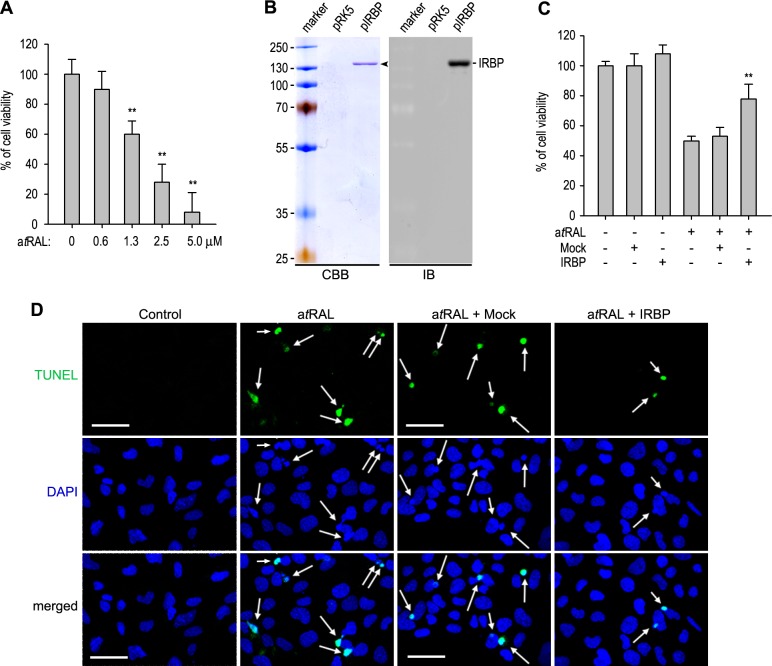
Interphotoreceptor retinoid-binding protein alleviated cell death induced by atRAL. (A) Viabilities of 661W cells incubated with the indicated concentration of atRAL in serum-free medium were determined by MTS assay. **P < 0.01 indicates significant differences between control and atRAL-treated cells. (B) Coomassie Brilliant Blue staining and IB analysis of IRBP in serum-free media of 293T-LC cells transfected with pRK5 or pIRBP. (C) Viabilities of 661W cells incubated with 0 or 1.8 μM atRAL in fresh medium or medium of 293T-LC cells transfected with pRK5 (mock) or pIRBP (IRBP). (D) TUNEL assay of 661W cells incubated with 1.8 μM atRAL as in (C). Nuclei were counterstained with 4′,6-diamidino-2-phenylindole (DAPI). TUNEL-positive nuclei were indicated with arrows. Scale bars: 50 μm.

