Figure 4.
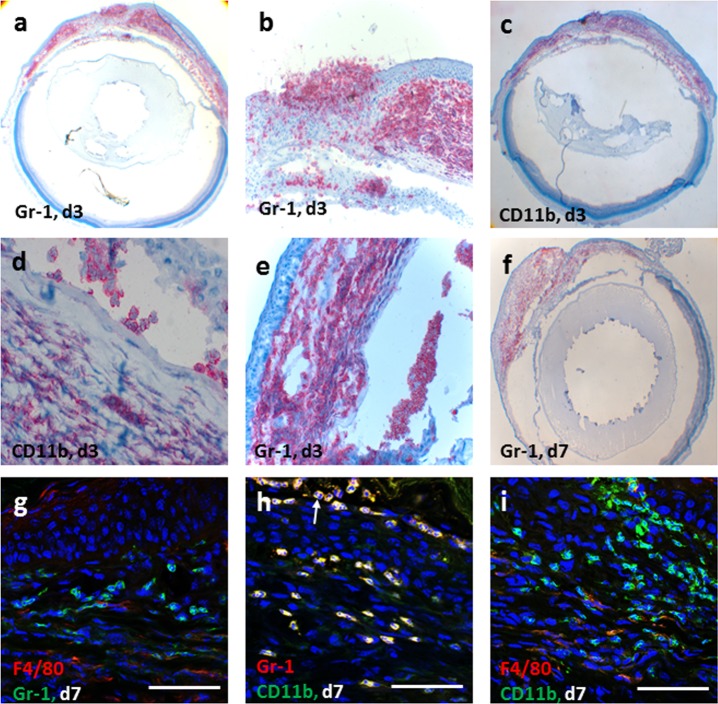
Infiltration of macrophages and neutrophils into corneal grafts in WT HSK mice. Corneal allografts stained with Gr1 and CD11b antibodies in early phase post corneal graft, day 3 (a–e) and day 7 (f) in HSK recipients show massive neutrophil (Gr1+) and macrophage (CD11b+) infiltration in the recipient cornea as well as in the donor corneal graft and anterior chamber of the eye. Dual staining of confocal microscopy images of day 7 grafts (g–i) show that majority of these cells are neutrophils (Gr1+CD11b+F4/80−) and macrophages (Gr1−CD11b+); white arrow indicating multilobed nucleus at day 7 post grafting (h). The F4/80+ cells (g, i) were infrequently observed cells in the inflammatory cell infiltrate and did not colocalize with Gr1+cells (g). Scale bars: 50 μm.

