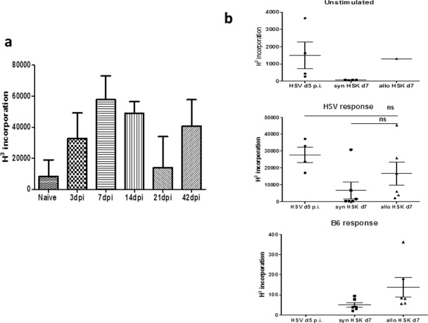
Figure 7.

Detection of anti–HSV-1 lymphocyte proliferation responses in the eye-DLN. (a) Lymphocyte proliferation from eye-DLN at different times after infection with HSV-1. Note consistent anti-HSV response even at the time-point when clinically the cornea is healed (i.e., day 42 dpi = time-point of corneal grafting procedure). (b) Lymphocyte proliferation responses to HSV antigen and B6 alloantigen day 7 post corneal allo- and syngraft (i.e., = day 49 pi). The response to HSV antigen after allografting procedure (middle panel) was equal to the lymphocyte response to HSV antigen 5 dpi in the ungrafted cornea. The difference between the allo- and syngraft response is not significant (see main text).
