Figure 2.
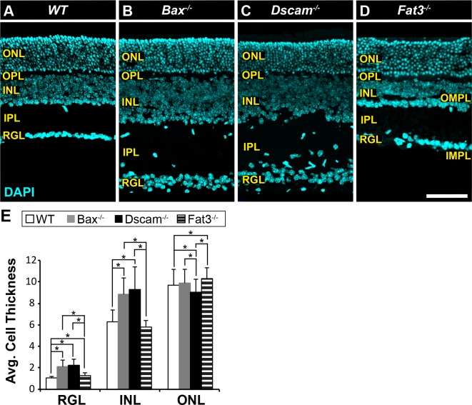
Mutant mouse strains used in this study. (A–D) Postnatal day 28 retina sections stained with DAPI. (A) WT retina. The stereotypic organization of the retina consists of three cellular laminae: ONL, INL, and RGL, separated by two plexiform layers: OPL and IPL. (B) Bax−/− retina. Increased thickness in the INL, IPL, and RGL due to a defect in developmental cell death. Lamination of the neural retina is maintained, except ectopic neural somas located in IPL. (C) Dscam−/− retina. Increased thickness in the INL, IPL, and RGL due to a defect in developmental cell death. Lamination of the outer retina is maintained while the inner retina (INL, IPL, and RGL) is highly disrupted. (D) Fat3−/− retina. Cellularity is maintained. Abnormal migration of neurons results in more cells found within the RGL and fewer in the INL. Two ectopic plexiform layers form. One in the INL and the other below the RGL, termed the OMPL and IMPL, respectively. (E) Cell thickness of each nuclear layer quantified; n ≥ 3 mice were used for each strain. Error bars: SD. A 1-way ANOVA was used to statistically analyze data for each cellular layer separately (RGL: P ≤ 6.61 × 10−52; INL: P ≤ 1.45 × 10−44; ONL: P ≤ 7.31 × 10⁻⁵). Tukey's pairwise comparison was used to compare each group. *Denotes statistical differences between groups. Scale bar: (D) 100 μm.

