Figure 8.
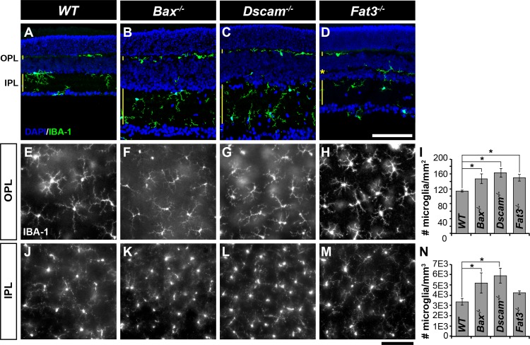
Microglia in mutant strains. (A–D) Postnatal day 28 retina sections stained with IBA-1 and DAPI. Microglia were found within neurite-containing laminae of retina: the OPL, IPL, and the OMPL of Fat3−/−mice. (E–H) Microglia within the OPL in whole retinas. (I) Graph illustrating density of microglia within the OPL. (J–M) Microglia within the IPL in whole retinas. (N) Graph illustrating density of microglia within the IPL. Within the OPL, a significant increase in microglia was observed in all models, when compared with WT. Within the IPL, a significant increase in microglia was observed in Bax−/− and Dscam−/− when compared with WT. n ≥ 3 mice was used for each strain per quantification. Error bars: SD. A 1-way ANOVA was used to statistically analyze the data (OPL: P ≤ 1.80 × 10⁻³; IPL: P ≤ 6.02 × 10⁻³). Tukey's pairwise comparison was used to compare between groups. *Denotes statistical differences. Scale bars: (D, M) 100 μm.

