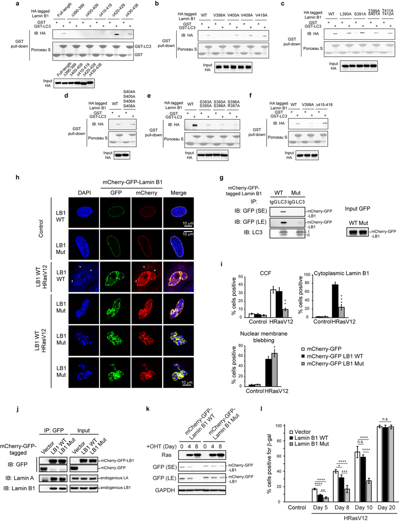
Extended Data Figure 9. Additional characterization of Lamin B1 substitution mutant.
a–f, Related to Fig. 5a, in vitro translated proteins were analyzed by GST-LC3B pulldown. g, LC3 IP in HEK293T cells transfected as indicated. The remaining interaction with the mutant is likely due to the endogenous Lamin B1 that interacts with LC3 and the mutant, as shown in j. h, i, IMR90 were imaged under confocal microscopy and quantified. Bars are the mean ± s.d.; n=4, over 200 cells; * P < 0.05, ** P < 0.005, *** P < 0.0001; unpaired two-tailed Student’s t-test. j, HEK293T transfected were analyzed by IP. k, ER:HRasV12 IMR90 were induced with OHT and harvested for immunoblotting. l, IMR90 were quantified for β-gal positivity. Bars are the mean ± s.d.; n=4, over 200 cells; * P < 0.05, ** P < 0.01, *** P < 0.001, **** P < 0.0001, n.s., non-significant; one-way ANOVA coupled with Tukey’s post hoc test.

