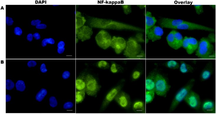
Fig 1. NF-κB/p65 nuclear localisation in differentiated THP-1 cells.
Cells were fixed, permeabilised, stained with anti-NF-κB/p65 rabbit monoclonal antibody and visualised with Alexa Fluor® 555 goat anti-rabbit IgG (green). The nuclei were counterstained with DAPI (blue). Microscopy images A. Inactive NF-κB/p65 proteins localisation in the cytoplasm of the non-stimulated THP-1 cells (top row) and B. Translocated NF-κB/p65 proteins into the nuclei of THP-1 cells following LPS stimulation (bottom row). Images were acquired for each fluorescence channel, using suitable TRITC and DAPI filters and a 63× objective. Scale bar: 10μm

