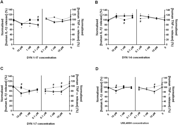
Fig 6. The modulation of DYN 1–17 and the N-terminal fragments on the release of IL-1β and TNF-α in LPS-stimulated THP-1 cells.
The LPS-stimulated THP-1 cells were treated with the N-terminal fragments of DYN 1–17 and U50,488H at 0.1 μM, 1 nM and 10 pM for 24 hr. The culture supernatants were collected and IL-1β and TNF-α release was measured using IL-1β and TNF-α AlphaLISA kit, respectively. The AlphaLISA signal was read using an Enspire-Alpha 2390 Multilabel Plate Reader. Non-stimulated THP-1 cells (NS) served as negative control. The release of IL-1β and TNF-α in each treatment group was normalised and expressed relative to LPS-stimulated control group. Data shown are the means ± S.E.M. of at least three independent experiments performed in triplicates. Statistical significance as denoted by * and # represent the IL-1β and TNF-α release between peptide-treated (solid-line) and LPS-stimulated control group or ML-190-treated group (dotted-line), respectively, p ≤ 0.05.

