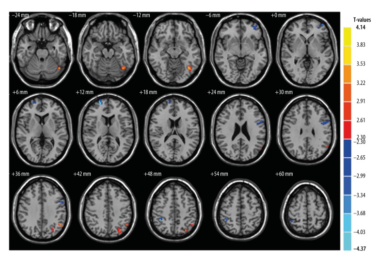
Figure 2.
Significant differences of WMV between the ON group and HCs. ON patients had significantly decreased WMV values in the brain regions of the left middle frontal gyrus, right superior frontal gyrus, left precentral gyrus and right inferior parietal lobule (blue areas) and significantly increased WMV values regions in ON were in the left fusiform gyrus, and left inferior parietal lobule (red areas). WMV,– white matter volume; ON – optic neuritis; HCs – health controls.

