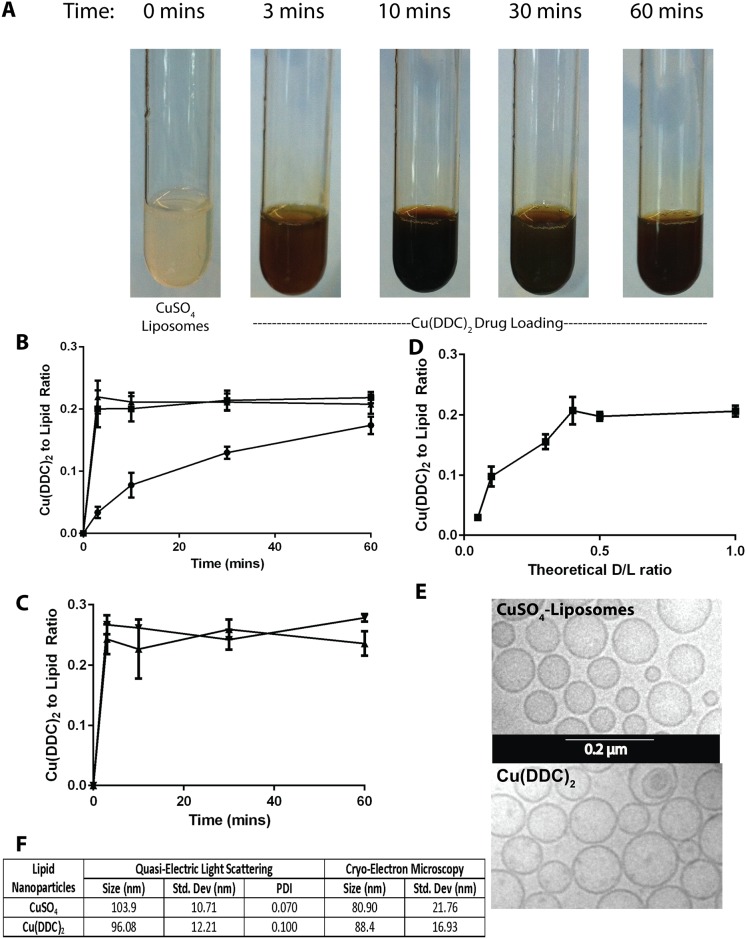
Fig 2. Diethyldithiocarbamate (DDC) loading into DSPC/Chol (55:45) liposomes prepared with encapsulated 300 mM CuSO4.
(A) Photograph of solutions consisting of DDC (5mg/mL) and added to CuSO4-containing DSPC/Chol (55:45) liposomes (20 mM liposomal lipid) over a 1 hour at 25°C. (B) Formation of Cu(DDC)2 inside DSPC/Chol liposomes (20 mM) as a function of time over 1 hour at 4(●), 25(■) and 40(▲)°C following addition of DDC at a final DDC concentration of (5 mM); Cu(DDC)2 was measured using a UV-Vis spectrophotometer and lipid was measured using scintillation counting. (C) Cu(DDC)2 formation inside DSPC/Chol (55:45) liposomes over time where the external pH was 7.4 (▲) and 3.5 (▼). (D) Measured Cu(DDC)2 as a function of increasing DDC added, represented as the theoretical Cu(DDC)2 to total liposomal lipid ratio; where the lipid concentration was fixed at 20 mM and final DDC concentration was varied. (E) Cryo-electron microscopy photomicrograph of CuSO4- containing DSPC/Chol (55:45) liposomes and the same liposomes after formation of encapsulated Cu(DDC)2. (F) Size of the CuSO4- containing liposomes and liposomes with encapsulated Cu(DDC)2 as determined by quasi-electric light scattering and cryo-electron microscopy; data points are given as mean ± SD.

