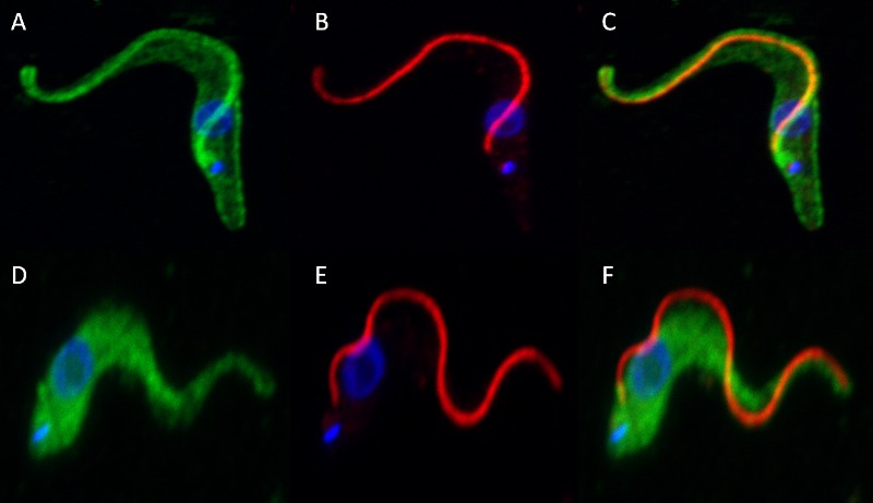
Fig 5. Confocal immunofluorescence microscopy showing localization of T. congolense calflagin in the presence and absence of calcium.

Maximum intensity projections of fixed T. congolense PCF were probed with mAb Tc6/42.6.4 and anti-para-flagellar rod protein in the presence (A, B, and C) and absence (D, E, and F) of calcium. Green: calflagin; Red: para-flagellar rod protein; Blue: DAPI/DNA.
