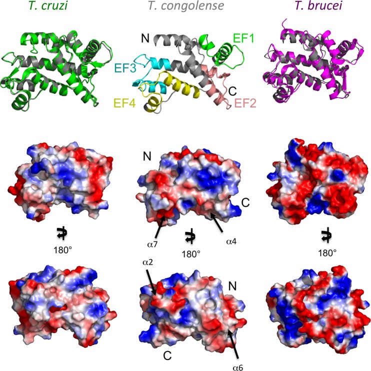
Fig 6. Structural characterization and surface analysis of T. congolense calflagin Modeled T. congolense calflagin (middle panel) was compared to the crystal structure of T. cruzi FCaBP (left vertical column) and to the NMR structure of T. brucei Tb24 (right vertical column).
(A) the predicted model of T. congolense calflagin (middle vertical column) exhibits four EF-hands motifs. The EF-hands (EF1, EF2, EF3 and EF4) are colored green, salmon, cyan, yellow, accordingly. Left panel: structural alignment of T. congolense (grey) over T. cruzi FCaBP (green). Right panel: T. congolense calflagin structure aligned over T. brucei Tb24 (magenta). (B) Surface representation of the three calflagin models overlapping panel A, showing exposed hydrophobic (gray), basic (blue) and acidic (red) respectively. (C) 180° rotation of B in the y-axis. Black arrows pointing towards putative epitopes and numbered according to their respective α-helix location.

