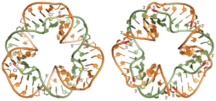
Figure 4.
Crystal structure of the self-assembling RNA nanotriangle. Views from both sides of the triangle plane are shown. The back view (left) reveals three Cl− ions (yellow spheres) bound at the Watson-Crick edge of A374 and C375. The terminal residues of all constituting oligonucleotides reside on one face of the triangle (front view, right). 5′ termini are highlighted in red. Atomic coordinates and structure factors have been deposited in the Protein Data Bank (PDB ID: 5CNR).

