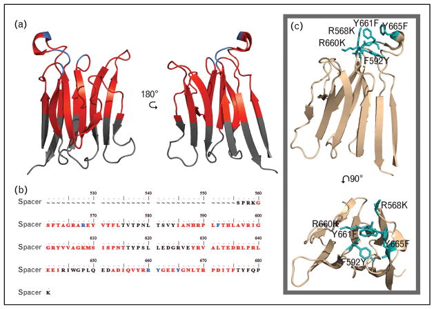
FIGURE 3.
Spacer domain sequence and structure (Protein Data Bank identity, 3GHM) with antibody antigenic regions marked, and gain of function (GoF) mutation. (a) Structure. (b) Sequence of the spacer domain with antigenic regions marked. Black text or regions designate no reported antibody exosite regions. Previously reported antibody binding residues are illustrated in blue. Red regions or text designates antibody exosite regions identified by conformational chemically linked peptides on scaffolds peptide technology. (c) ADAMTS13 spacer domain structure with the GoF mutations in teal.

