Figure 6.
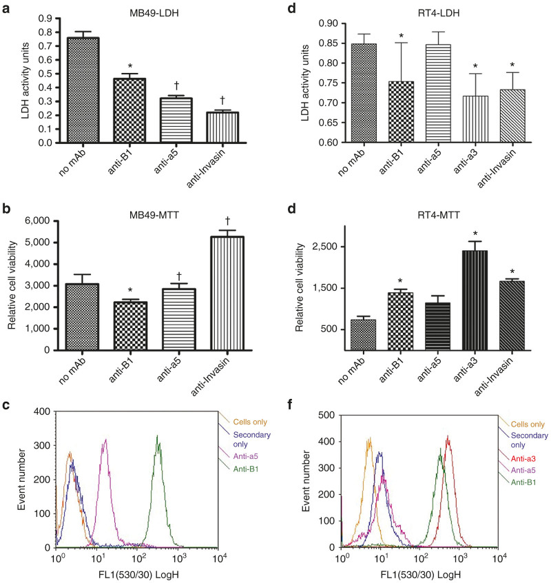
Integrin-specificity of VAX-IP. (a–c) The ability of VAX-IP to stimulate LDH release (a) and cell killing (b) in MB49 cells was assessed in the presence of function blocking anti-mouse β1 or α5 integrin antibodies as well as with VAX-IP minicells preincubated with the Invasin function blocking monoclonal antibody mAb3A2. The relative expression levels of both β1 and α5 in MB49 is demonstrated by flow cytometry using the same function blocking antibodies as primary detection reagents (c). (d–f) The ability of VAX-IP to stimulate LDH release (d) and cell killing (e) in RT4 cells was assessed in the presence of function blocking anti-human β1, α3, or α5 integrin antibodies as well as with VAX-IP minicells preincubated with the Invasin function blocking monoclonal antibody mAb3A2 or an irrelevant antibody control. The relative expression levels of β1, α3, and α5 in RT4 is demonstrated by flow cytometry using the same function blocking antibodies as primary detection reagents (f). * denotes P ≤ 0.05; † denotes P ≤ 0.005.

