Figure 2.
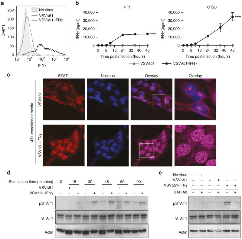
Virus-expressed IFNγ is secreted and functional. (a) Flow cytometry histogram showing the intracellular IFNγ staining of 4T1 cells uninfected or infected with VSVΔ51 or VSVΔ51-IFNγ for 18 hours at a multiplicity of infection (MOI) of 0.01. (b) IFNγ concentrations determined by enzyme-linked immunosorbent assay of the supernatants of 4T1 and CT26 cells infected with VSVΔ51 or VSVΔ51-IFNγ at an MOI of 0.01 for various periods of time. Results represent values obtained from duplicate infections. ***P < 0.001 (two-way analysis of variance). (c) Confocal microscopy pictures of 4T1 cells stained with a STAT1-specific antibody and 4’,6-diamidino-2-phenylindole. Supernatant from 4T1 cells uninfected or infected for 24 hours at an MOI of 3 was collected and transferred onto fresh 4T1 cells for 60 minutes upon virus removal (virus-cleared conditioned media). The gray boxes indicate the region that is enlarged in the right panels. (d) and (e) Western blot analysis of phosphorylated-STAT1 (pSTAT1), STAT1 and actin. Virus-cleared conditioned media was transferred onto fresh 4T1 cells for various periods of time. (e) For half the conditions, the supernatant was incubated with an IFNγ-blocking antibody prior to a 20-minute incubation with the fresh cells.

