Figure 4.
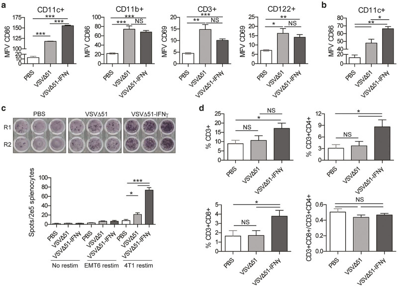
IFNγ-expressing virus induces greater activation of dendritic cells. (a) 4T1 lung tumor-bearing mice were treated intravenously with a single dose of virus or phosphate-buffered saline (PBS) (n = 4). Twenty-four hours post-treatment, splenocytes were harvested and stained for CD11c, CD11b, CD86, CD3, CD122, and CD69 24 hours post-treatment. Samples were analyzed by flow cytometry. The graphs show the mean fluorescence values. Data are representative of two different experiments. (b) 4T1 fat-pad tumor-bearing mice were treated intratumorally with one dose of virus or PBS. Twenty-four hours post-treatment, the splenocytes were stained for CD11c and CD86. (c) IFNγ ELISPOT of splenocytes restimulated ex vivo with 4T1 tumor cells. 4T1 fat-pad tumor bearing mice were treated with PBS, VSVΔ51, or VSVΔ51-IFNγ. Ten days post-treatment, splenocytes were harvested and the ELISPOT was performed. R1 and R2 are two replicates from the same mouse. The bar chart represents the counts obtained for each group. (d) Single cell suspensions were obtained from tumors of the same mice and stained for CD3, CD4, and CD8. Samples were analyzed by flow cytometry. The graphs show the percentage of the cells within the tumor that were positive for the different markers. NS: P > 0.1, *P < 0.1, **P < 0.01, ***P < 0.001 (unpaired one-tailed T-test).

