
Figure 3.
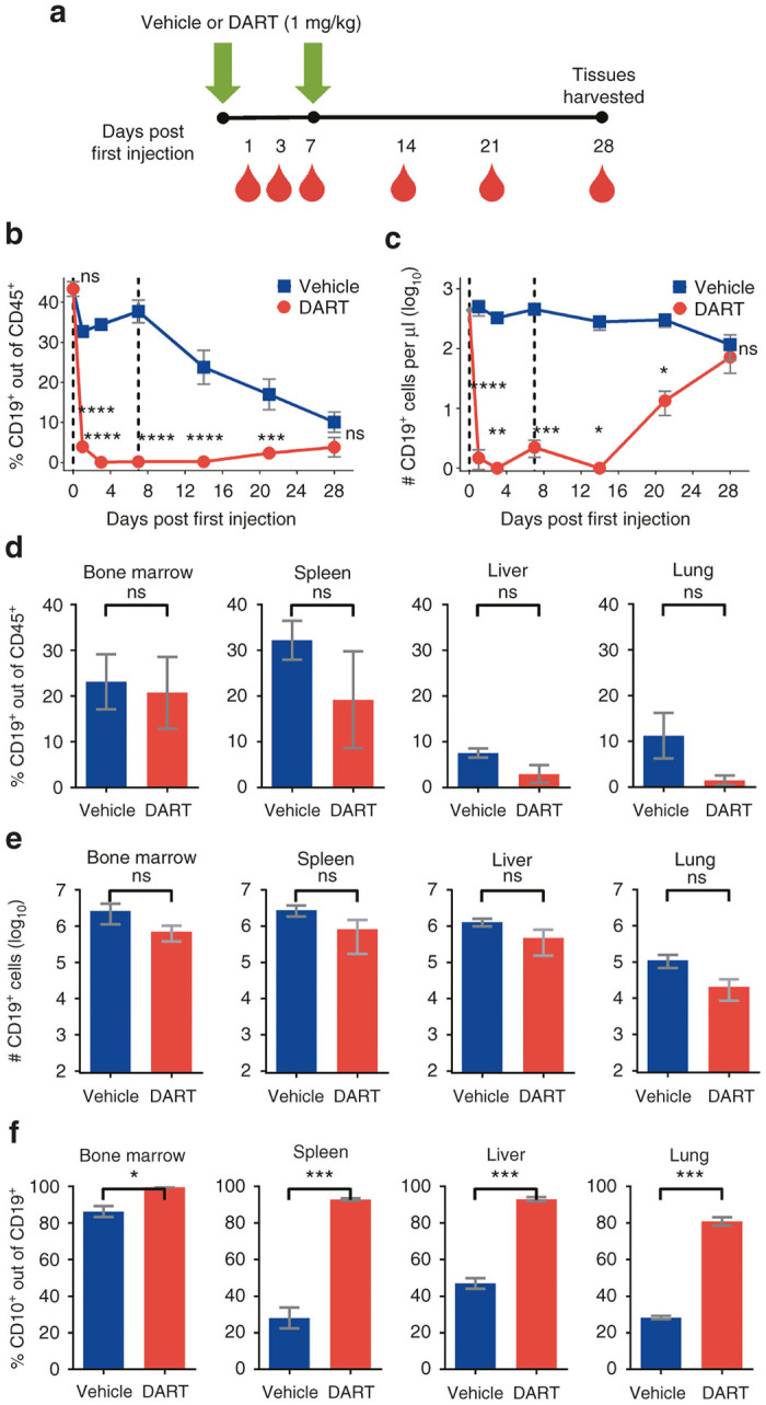
Immature human CD19+ B cells regenerate in NSG/BLT mice after CD19xCD3 DART protein administration. (a) Experimental outline. NSG/BLT mice were administered CD19xCD3 (n = 3, 1 mg/kg i.v.) or vehicle (n = 3) at day 0 and day 7. Peripheral blood (PB) was collected and analyzed at days 0, 1, 3, 7, 14, 21, and 28; tissues were harvested and analyzed at day 28. (b) Percent CD19+ cells out of CD45+ cells and (c) number CD19+ cells per microliter detected in PB by flow cytometry (vehicle group in blue; CD19xCD3 group in red). (d) Percent CD19+ cells out of CD45+ cells and (e) number CD19+ cells detected in tissues by flow cytometry. (f) Percent immature CD10+ cells out of CD19+ cells detected in tissues by flow cytometry. (Mean ± SEM plotted. P values were calculated in b and c by repeated-measures two-way analysis of variance with Sidak’s multiple comparisons test, comparing vehicle versus CD19xCD3. P values were calculated in d–f by unpaired t-test, comparing vehicle versus CD19xCD3. ns, P > 0.05; *P < 0.05; **P < 0.01; ***P < 0.001; ****P < 0.0001. Gray diamond indicates data from age-matched NSG/BLT mice, n = 4.)