
Figure 8.
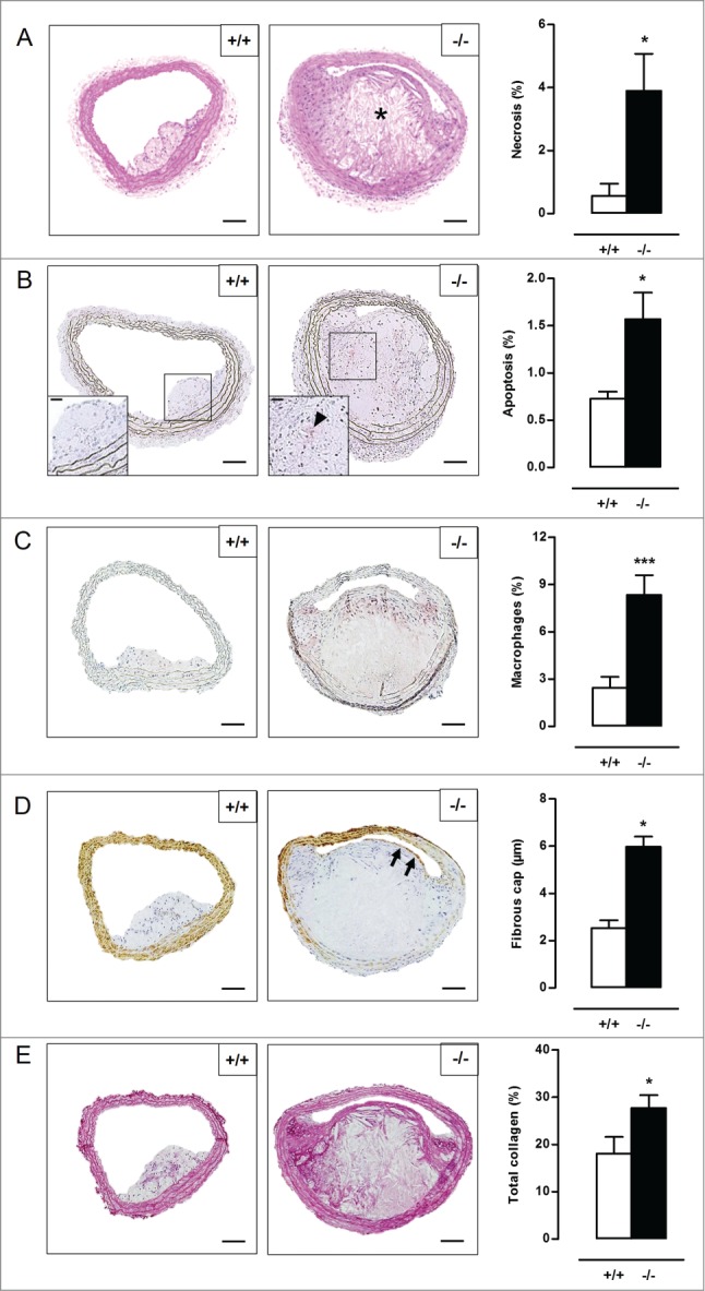
Defective VSMC autophagy accelerates atherogenesis after 10 wk of western-type diet. (A) Atg7+/+Tagln-Cre+ apoe−/− (+/+) and Atg7F/FTagln-Cre+,apoe−/− (−/−) mice (n = 16) were fed a western-type diet for 10 wk. Sections of the brachiocephalic artery were stained with H&E to quantify plaque size and percentage of necrosis. (*, P<0.05; Student t test). (B to D) Consecutive sections were immunostained for cleaved CASP3 (B), LAMP2 (C) and ACTA2 (D) to measure the percentage of apoptosis (indicated by a black arrowhead in the high-power photograph of the boxed area in the left corner of each image), percentage of macrophages and fibrous cap thickness (indicated by black arrows), respectively (*, P < 0.05; Student t test (B); ***, P < 0.001; Student t test (C); *P < 0.05; n = 10 measurements/mouse, Repeated Measure (D)). (E) Consecutive sections were stained with Sirius red to quantify total collagen. (*, P < 0.05; Student t test). *, necrotic core. Scale bar: 100 µm.