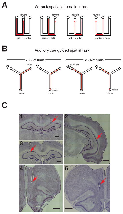
Figure 1. Behavior Paradigms and Recording Locations.
(A) W-track. Animals had to learn to alternate between the three arms of the W-track for reward.
(B) Y-track. In each trial initiated by poking in the home well, animals had to learn to visit the “silent well” when no sound was presented (75% of trials) and the “sound well” when the target sound was presented (25% of trials).
(C) Histology illustrating recording locations. Nissl stained coronal sections showing electrode tracks and lesion location. (C1) W-track: dorsal CA1, (C2) W-track: intermediate CA1, (C3) Y track: dorsal CA1, (C4) W-track: medial PFC, (C5) Y track medial PFC. Scale bars are 1 mm.

