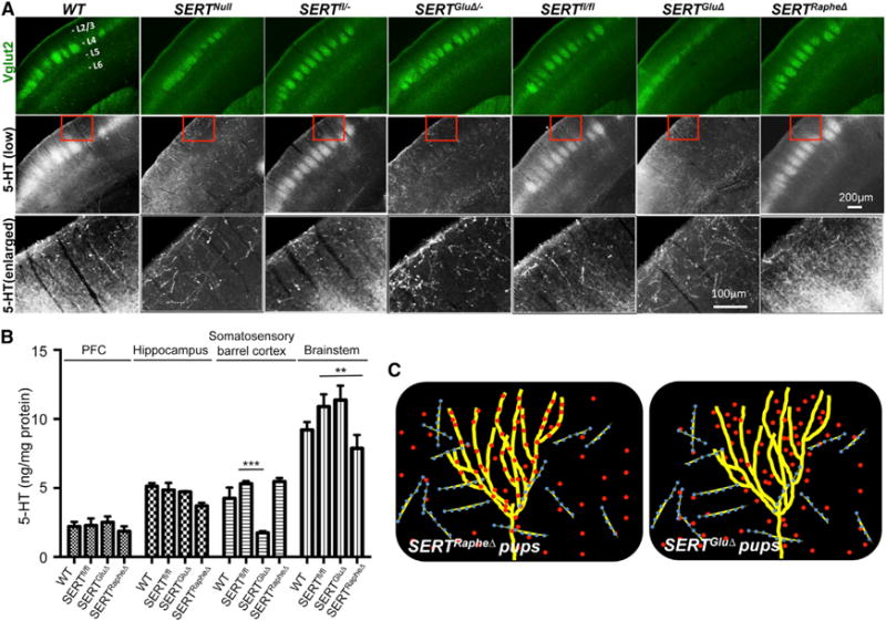
Figure 3. Requirement of SERT Expression in TCAs for 5-HT Uptake in the Barrel Cortex.

(A) Double immunohistochemistry for Vglut2 and 5-HT on coronal sections of the somatosensory cortex of P7 mice. The positions of cortical layers are indicated. Third row images show an enlarged view of 5-HT immunohistochemistry in areas outlined by red boxes on images in the second row. Note two types of 5-HT+ fibers in WT and control mice: fibers that colocalized with Vglut2+ TCA patches at layer IV (L4, strongly) and layer VI (L6 weakly), and Vglut2-negative fibers, which were thicker, highly punctated, and randomly distributed. 5-HT staining in Vglut2+ TCAs, not the thicker fibers, was abolished in SERTGluΔ and SERTGluΔ/ and SERTNull mice. 5-HT staining in Vglut2+ TCAs in SERTRapheΔ mice was indistinguishable from the control littermate and WT mice. Cortical sections from mutant and control littermate mice were always stained in parallel. For each of the brain samples, alternate sections were analyzed by double immunostaining of Vglut2 and SERT, and data are presented in Figure S4.
(B) HPLC analysis of 5-HT concentrations in brain tissues collected from P7 mice. Values from SERTfl/fl littermates of SERTGluΔ and SERTRapheΔ mice were pooled and compared to that of WT, SERTGluΔ, and SERTRapheΔ mice (n = 4, mean ± SEM, **p < 0.01, ***p < 0.001, one-way ANOVA). SERT protein abundance in each of the brain samples was analyzed by western blotting shown in Figure 2C.
(C) Model for SERT expressed in TCAs and raphe afferents during barrel map development: Left, in SERTRapheΔ mice, TCA-expressed SERT clears 5-HT from the sensory cortex for barrel map development. Right, in SERTGluΔ mice, trophic 5-HT accumulates around growing TCAs despite SERT expression in raphe afferents and impairs barrel map formation. Yellow lines denote TCAs, blue/yellow lines denote raphe neuron axons, and red dots denote 5-HT.
