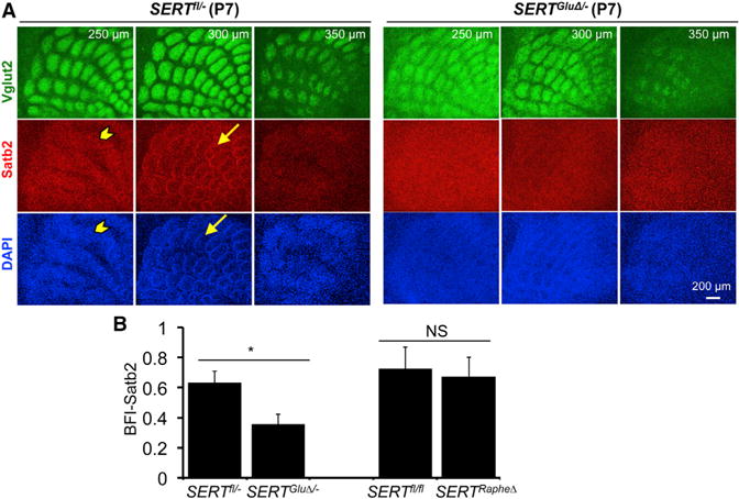
Figure 6. SERT Expressed in TCAs Governs Multiple Spatial Organizations of Satb2+ Neurons in the Somatosensory Cortex.

(A) Serial sections through the PMBSF of P7 mice labeled by double immunostaining of Vglut2+ TCAs and Satb2+ cell nuclei, with DAPI counterstain. The distance from the pial surface is indicated. In control littermates (SERTfl/−), Satb2+ cells, as well as DAPI-labeled nuclei, displayed two distinct patterns: stripes (arrowhead) located ~250 μm from the pial surface and barrels (arrow) located below the stripe structure. Both Satb2+ patterns were largely absent in SERTGluΔ/− mice. Impaired spatial organization of other neuronal markers in P7 and mature barrel cortex of SERTGluΔ/− mice is shown in Figure S7.
(B) Evaluation of Satb2 barrel structure by calculating BFI of Satb2 immunohistochemistry in P7 PMBSF (n = 5, mean ± SEM, *p < 0.05, Student’s t test).
