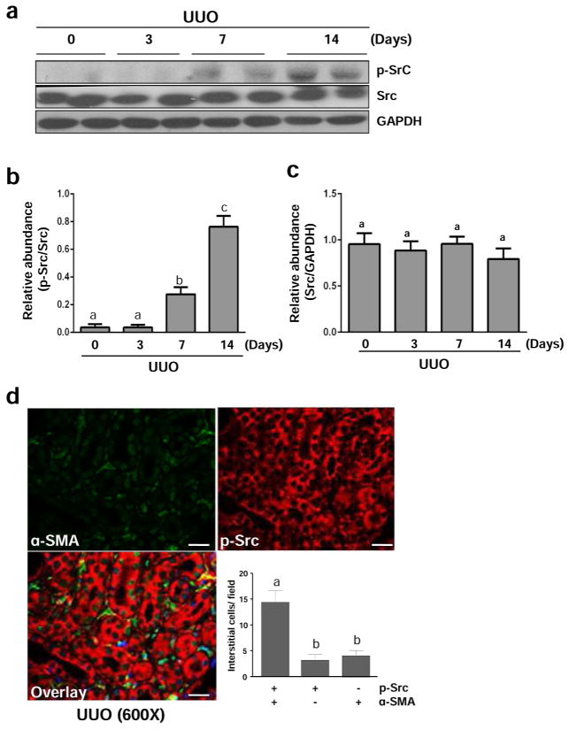
Figure 5. Src is activated in the kidney after UUO injury.
The left ureter was ligated for 3, 7, and 14 days. The kidneys were taken for immunoblot analysis of phospho-Src (Tyr-416) (p-Src), Src, GAPDH as indicated (a). Representative immunoblots from 3 experiments are shown. Expression levels of p-Src and Src were quantified by densitometry and normalized with GAPDH as indicated (b, c). Photomicrographs illustrate immunoflurecent staining of kidney tissue taken from the kidney subjected to UUO for 7 days with the antibodies to p-Src and α-SMA (600 x). The number of interstitial cells expressing p-Src, α-SMA, or p-Src + α-SMA were accounted respectively (d). Data are represented as the mean ± SEM (n=6). Bars with different letters (a–c) are significantly different from one another (P<0.05). Scale bar=20 μM.

