Fig. 1.

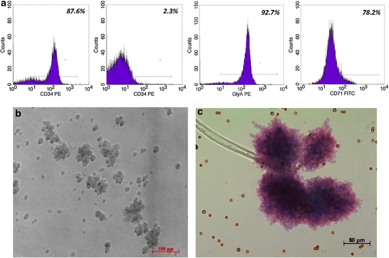
Flow cytometry showing differentiation of CD34+ cells to erythroid progenitors (a) and the cluster-like basophilic erythroblast morphology of erythroid cells on day 7 of differentiation (b) and the colony-forming hematopoietic cell units (CFU-E) (c). CD34+ hematopoietic stem cells were purified from umbilical cord blood mononuclear cells using Dynabead magnetic separation to a purity of 87.6 %. After the 15 day single-phase ex vivo expansion and differentiation, the cell population was 92.7 and 78.2 % CD235a+ and CD71+, respectively with minimal CD34 positivity. This confirmed our differentiation and provided erythroid cells for down-stream experiments (a). During the expansion and differentiation of HSCs to erythroid cells, the cells formed cluster-like colonies before separating into single cells in suspension, which was typical of terminal differentiation (b). The hematopoietic colony formation assay was performed to confirm the stem-like behaviour of the Dyna-bead selected CD34+ HSCs prior to expansion and differentiation. The colony-forming unit- granulocyte, erythroid, monocyte, megakaryocyte (CFU-GEMM) semi-solid cultures were grown for 14 days and stained for colony counting (c)
