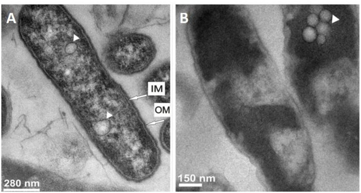
FIGURE 1.
Ultrastructural changes in Legionella dumoffii cells after treatment with Galleria mellonella apolipophorin III. Transmission electron micrographs of (A) untreated bacteria and (B) treated bacteria with 0.4 mg/ml ApoLp-III. The presence of vacuoles is indicated by arrowhead. IM, Inner membrane; OM, Outer membrane (Source: Palusinska-Szysz et al., 2012).

