
Fig. 2.
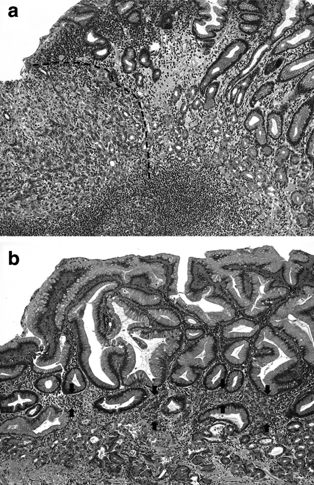
Growth patterns at the edge of the intramucosal component. The expansile pattern demonstrates destructive invasion with a relatively well-defined margin (indicated by the dotted line) at the advancing edge (a). In the permeative pattern, neoplastic cells infiltrate between the normal pits or glands in the middle layer of the lamina propria, with no recognizable margin to the growth (b). Neoplastic cells are indicated by arrows