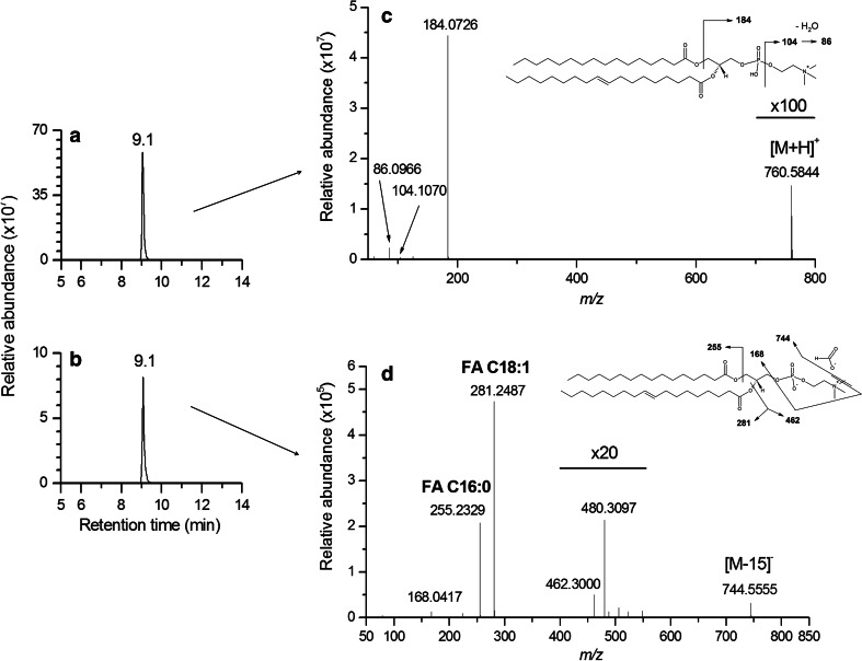
Fig. 4.
Extracted ion chromatograms of the PC34a:1 lipid species. The [M + H]+ ion at m/z 760.5844 (a), and the [M − H + CHO2]− ion at m/z 804.5778 (b) were detected in the same retention time range (9.1 min ± 10 s), in the positive and negative ion mode, respectively. The CID mass spectrum of the protonated [M + H]+ ion confirmed the presence of the phosphocholine polar head (c), whereas the CID mass spectrum of the formate adduct [M − H + CHO2]− ion enabled the detection of the two FA C18:1 (m/z 281.2487) and FA C16:0 (m/z 255.2329) associated fatty acids (d), leading to an improved structural characterization of this lipid species

